Thursday, 30 January 2014


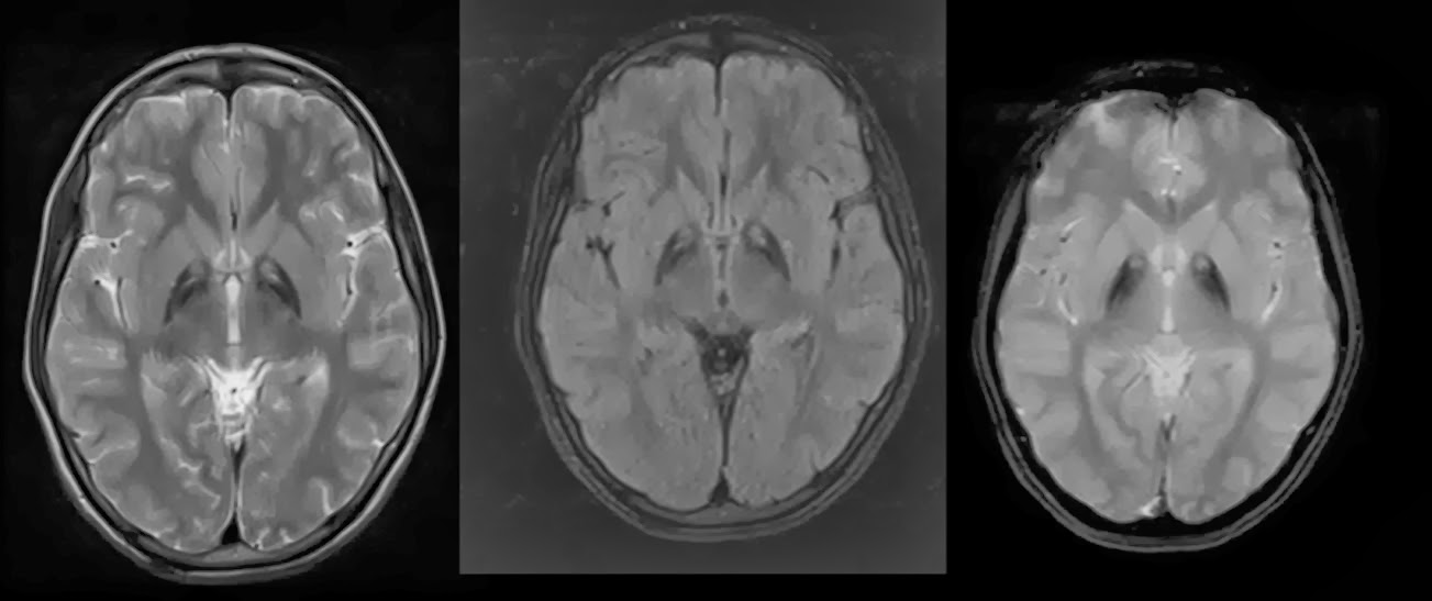
Atrocities of two German neuropathologists Julius Hallervorden and Hugo Spatz during Second World War are notorious. The eponymous term Hallervorden –Spatz syndrome is disfavoured and neurodegeneration with brain iron accumulation (NBIA) is encouraged.
Ante mortem diagnosis of this rare condition is made possible with the use of MRI examination. Two distinct clinical presentations are described one is classical form and another is atypical variant based on the age of onset. Both the forms are progressive with poor prognosis.
Patients present in early age with progressive gait impairment, rigidity of all limbs, dysarthria and mental retardation. 
MRI findings are striking and reflect the pathology. Preferential deposition of the iron in globus pallidus will result in its destruction. The MRI appearance is described as eye of the tiger appearance. This sign is seen in both T2W and FLAIR imaging. Foci of T2 Hyper intensity noted with in the area of T2 Hypo intensity. Hypo intensity reflecting the iron deposition and the hyper intensity reflecting the gliosis changes.
Ref:

Wednesday, 8 January 2014


All the cases of omental infarcts have been described in the last fifteen years only. These are increasingly recognised as an important cause of the right lower quadrant abdominal pain. Two factors contribute to the increased recognition of this entity. One is the availability of CT scanning technology and the other is epidemic of obesity.


Commonly the severity of the pain is disproportionate to the clinical signs. It is almost always located in the right iliac fossa in close association to the ascending colon and hepatic flexure. CT imaging is highly specific for omental infarcts. A cake like high attenuation fat density mass centered in the omentum is typically observed. If the adjacent bowel wall is thickened it is very minimal and pathology is primarily located in the omentum.
Omental infarcts are classified as primary and secondary types. If an aetiology is identified it is called as secondary and if no cause could be found it is called as primary.
Omental infarcts and epiploic appendagitis may have similar appearance, but omental infarct is a right sided pathology where as epiploic appendagitis is a left sided pathology. When it is not possible to differentiate an umbrella term intrabdominal focal fat inflammation can be used with no harm.

The importance in recognising these entities is to prevent unnecessary surgery. Most of the cases of omental infarcts get well with conservative management but when there is detoriation in the patient condition surgery and necrosectomy can be performed with out hesitation. In this patient there is nonrotation of the small bowel with midgut volvulus and this can be precipitating factor for omental infarct 

Ref:


Wednesday, 1 January 2014


Osteopetrosis is a developmental bone abnormality radiologically characterised by generalised increase in bone density. Bone appears chalky white and is brittle. Paradox it may seem but they are more prone for fractures like osteoporotic bones.

Heinrich Ernst Albers-Schonberg of Hamburg (Germany) first described this condition. He was one of the pioneer radiologist and many radiographic techniques used in day to day practice were developed by him. He is well known for is early reports of radiation induced damage of the rabbit ovaries in 1903.

Excessive resorption of the bone causes osteoporosis, similarly defective bone resorption leads to osteopetrosis. At least eight subtypes of osteopetrosis are described. Tarda variant of osteopetrosis is encountered in adults and have reasonably good prognosis. Mode of inheritance is autosomal dominant. More malignant varieties are encountered is early age group and show autosomal recessive inheritance.

Long bones show generalised increased density and undertubulation of the bones resulting in the Erlenmeyer flask deformity. Joint is spared. Sclerosis of the vertebral endplates results in sandwich vertebra. Failure of the osteoclasts to remodel the bone result in endobone formation or bone with in bone appearance. 

Ref:

1.http://jama.jamanetwork.com/article.aspx?articleid=381625-The 'Bone Within a Bone'David H. Frager, MD; Kakarla Subbarao, MD

2. http://hmg.oxfordjournals.org/content/10/25/2861.full- perspective from genetics

3. http://www.ijri.org/article.asp?issn=0971-3026;year=2003;volume=13;issue=4;spage=387;epage=388;aulast=Phatak- case report and review

4. http://www.ajnr.org/content/21/6/1110.long-  cranial manifestations

5. http://dentistry.tbzmed.ac.ir/joddd/index.php/joddd/article/viewFile/398/261 - case reports and review

6. http://bjr.birjournals.org/content/79/940/359.full- case report from BJR